Digitized Radiological Diagnostics
The Ruesch Clinic provides its patients with specialized medical teams and high-profile equipment making the facility a benchmark for diagnostics in Southern Italy.
General Info
The radiography can be either the radiographic image or the actual radiogram or the radiographic technique used in obtaining the radiogram. Radiology is the branch of medicine that deals with radiography.
Radiography is a technique of medical diagnosis that is mainly based on the use of X-rays and their braking effect due to the interaction that occurs between the material (that is, our body) and radiation. X-rays – which are a beam of photons – interact from a source and a receptor between which the matter to be examined is interposed. The atoms of this biological body prevent some photons from reaching the receptor by “blocking” and thus providing an image of the body examined so to speak “in negative”, all those photons that are not absorbed by the receptor are impressed on the film.
The radiography was invented by Wilhelm Conrad Rontgen in 1985 and over the decades has undergone many changes and evolutions that have led today to the use of computerized radiography in medicine.
Insights
Why are radiographs made?
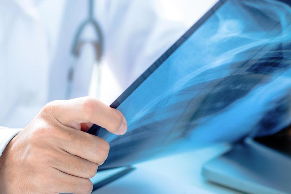
To obtain precise radiological images, the radiology technician will have to place the patient on a special table or on a screen, where he will have to remain immobile for a few moments. In fact, X-rays remember to be ionizing radiations that do not give any effect when they cross the body, but even a slight movement could compromise the focus of the plates. However, the X-ray beam penetrates the human body and is absorbed differently depending on the part taken into consideration, attenuating and then being absorbed by the receptor and then reaching the detection system.
Nowadays, with the advent of digital radiography, the photographic film that was previously a fundamental part in the development of radiographs has been replaced by radiographic cassettes that present in themselves photostimulable substances and which are inserted into special readers that return to the computer a good image quality obtained from the analysis.
After completing this procedure, the radiology technician will pass to the radiologist who will take care to analyze the cassettes and provide a report.
To date, radiography is still one of the most important techniques used especially in emergency cases because it is a medical investigation with a particularly simple and fast development and also relatively cheap, as well as being able to provide a clear and detailed image in the study of the pathologies of the human body.
Who can make an X-ray? Are there any health risks?
There are no special restrictions for the public who can use a radiology exam. Some tricks, however, must be: women of childbearing age, for example, must be certain that they can rule out a pregnancy in place, as X-rays can be very harmful to the fetus. In this regard, in fact, the radiography is not recommended for pregnant women and is performed only in cases of extreme urgency and only if it is not possible to replace it with other equally specific tests that do not use x-rays.
Radiography is not a painful practice: the ionizing rays do not cause any pain when they pass through the patient’s body. But in any case X-rays have a biological effect on the tissues examined that they cross, in fact, although the radiation for a single radiograph is in any case very low density, each radiological examination must always be justified by precise diagnostic requirements such as urgency or the diagnosis of bone pathologies such as osteoarthritis, osteoporsi or the investigation of deformations of the vertebral column such as scoliosis, kyphosis and lordosis and so on.
Radiography Naples
We provide the best diagnostic services for all patients, also from neighboring cities, who want to reserve the best specialists and the best technologies for their health care.